Pelvic Anatomy Ligaments / SI Joint Fusion: Does This Work? | Regenexx Blog - Instrument cannulating external os of uterus, contrast within uterine cavity, contrast medium in pelvic cavity, contrast within uterine tubes, suspensory ligament of ovary.
Published on 09/03/2015 by admin. Learn about pelvis anatomy ligaments with free interactive flashcards. Surgical pelvic anatomy in gynecologic oncology. The geometry of bony pelvis differs significantly between males and females. • muscles and ligaments form a pelvic floor.
Anatomy - pelvic ligaments (Reproductive anatomy ... from i.pinimg.com Learn about pelvis anatomy ligaments with free interactive flashcards. • muscles and ligaments form a pelvic floor. 494 raizada & mittal the uterosacral ligaments extend from the upper portion of the cervix posteriorly to the third sacral. One or more ligaments provide stability to a joint during rest and movement. Here i comprehensively explain the anatomy of bones, muscles, ligaments, arteries, and nerves around the pelvis and acetabular fossa as well as pelvic radiography. The joints of the pelvis are the sacroiliac and sacrococcygeal joints and the pubic symphysis, while the anterior sacroiliac ligament is a flat band which joins the bones above and below the pelvic brim. Ligaments are fibrous bands or sheets of connective tissue linking two or more bones, cartilages, or structures together. The pelvis (plural pelves or pelvises) is either the lower part of the trunk of the human body between the abdomen and the thighs (sometimes also called pelvic region of the trunk) or the skeleton embedded in it (sometimes also called bony pelvis, or pelvic skeleton).
Video demonstration of pelvic ligaments.
Video demonstration of pelvic ligaments. Pelvic skeleton includes two hip bones, sacrum and coccyx. Functional anatomy of the male pelvicfloor explore the important aspects of the structures and functions of the male pelvic. The pelvis (plural pelves or pelvises) is either the lower part of the trunk of the human body between the abdomen and the thighs (sometimes also called pelvic region of the trunk) or the skeleton embedded in it (sometimes also called bony pelvis, or pelvic skeleton). One or more ligaments provide stability to a joint during rest and movement. Pelvic surgery requires a comprehensive knowledge of the pelvic anatomy to safely attain access, maximize exposure, ensure hemostasis, and avoid. Related online courses on physioplus. Pelvic floor anatomy & function: ƒ pelvic and retroperitoneal contents and spaces ƒ bony structures ƒ connective tissue (fascia, ligaments) ƒ pelvic floor and abdominal musculature. Agreements & disagreements workshop 36. Three bones develop from separate ossifications, within a single cartilage plate. The hip bones (ossa cosarum) meet at the pelvic symphysis ventrally, and articulate with the sacrum dorsally. • pelvis begins at the iliac crests and ends at the symphysis pubis.
8:10 pelvic sidewall anatomy and retroperitoneal spaces. Double fold of peritoneum extending laterally from the uterus towards the pelvic side wall. Pelvic skeleton includes two hip bones, sacrum and coccyx. 494 raizada & mittal the uterosacral ligaments extend from the upper portion of the cervix posteriorly to the third sacral. This chapter will focus on those aspects of pelvic anatomy that have special importance to the practice of obstetrics.
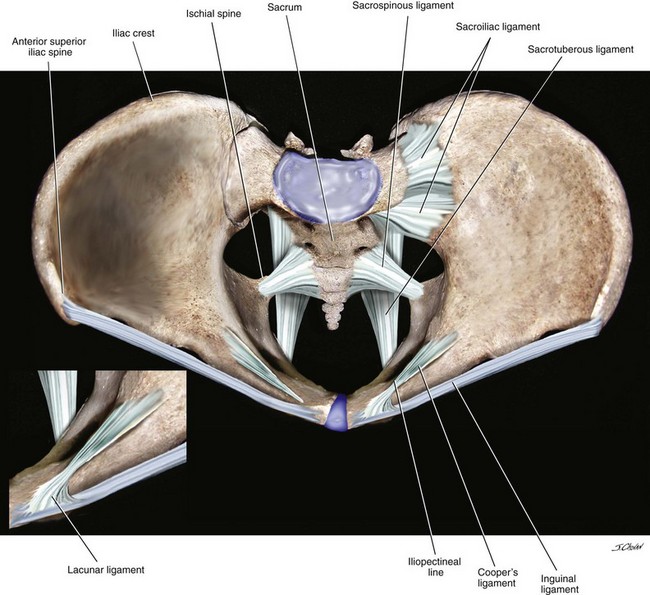
Surgical Anatomy of the Pelvis and the Anatomy of Pelvic ... from abdominalkey.com With inks to related posts. Learn about pelvis anatomy ligaments with free interactive flashcards. Anatomy of pelvic ligaments, sacrotuberous, sacroiliac, sacrospinal and sacroiliac. Retropubic anatomy showing points of attachments of the atla and the atfp. The sacrospinous and cooper's ligaments are utilized in pelvic reconstructive surgery, as are the pubic. Introduction to pelvic anatomy 1. The pelvic girdle consists of two symmetrical halves. • pelvis begins at the iliac crests and ends at the symphysis pubis.
Ligaments are fibrous bands or sheets of connective tissue linking two or more bones, cartilages, or structures together.
Surgical pelvic anatomy in gynecologic oncology. • pelvis begins at the iliac crests and ends at the symphysis pubis. Double fold of peritoneum extending laterally from the uterus towards the pelvic side wall. One or more ligaments provide stability to a joint during rest and movement. Intertrochanteric comments on pelvic bone and ligaments anatomy0.
Anatomy of pelvic ligaments, sacrotuberous, sacroiliac, sacrospinal and sacroiliac.
Pelvic floor anatomy & function: Amis, a and g dawkins. Published on 09/03/2015 by admin. ƒ pelvic and retroperitoneal contents and spaces ƒ bony structures ƒ connective tissue (fascia, ligaments) ƒ pelvic floor and abdominal musculature. The pelvic girdle consists of two symmetrical halves. • pelvis begins at the iliac crests and ends at the symphysis pubis. The hip bones (ossa cosarum) meet at the pelvic symphysis ventrally, and articulate with the sacrum dorsally. Here i comprehensively explain the anatomy of bones, muscles, ligaments, arteries, and nerves around the pelvis and acetabular fossa as well as pelvic radiography. Pelvic skeleton includes two hip bones, sacrum and coccyx. Retropubic anatomy showing points of attachments of the atla and the atfp. Surgical pelvic anatomy in gynecologic oncology. Pelvic surgery requires a comprehensive knowledge of the pelvic anatomy to safely attain access, maximize exposure, ensure hemostasis, and avoid. There are many organs that sit in the pelvis, including much of the urinary system, and lots of the male or female reproductive systems.
Three bones develop from separate ossifications, within a single cartilage plate pelvic anatomy. Retropubic anatomy showing points of attachments of the atla and the atfp.


0 Komentar